https://pubmed.ncbi.nlm.nih.gov/33093460/
Cell-penetrating peptide-mediated cell entry of H5N1 highly pathogenic avian influenza virus
- PMID: 33093460
- PMCID: PMC7582914
- DOI: 10.1038/s41598-020-74604-w
H5N1 highly pathogenic avian influenza virus (HPAIV) poses a huge threat to public health and the global economy. These viruses cause systemic infection in poultry and accidental human infection leads to severe pneumonia, associated with high mortality rates. The hemagglutinin (HA) of H5N1 HPAIV possesses multiple basic amino acids, as in the sequence RERRRKKR at the cleavage site; however, the role of this motif is not fully understood. Here, we showed that a 33-amino acid long peptide derived from HA of H5N1 HPAIV (HA314-46) has the potential to penetrate various cells and lung tissue through a sialic acid-independent endocytotic pathway. Mutant peptide analyses revealed that the cysteine residue at position 318 and multiple basic amino acids were essential for the cell-penetrating activity. Moreover, reassortant viruses possessing H5 HA could enter sialic acid-deficient cells, and virus internalisation was facilitated by cleavage with recombinant furin. Thus, our findings demonstrate that the HA314-46 motif exhibits cell-penetrating activity through a sialic acid-independent cell entry mechanism.
Introduction
Influenza A viruses are responsible for the seasonal epidemics and occasional pandemics in humans and animals1. These viruses have a segmented, single-stranded, negative-sense RNA genome within the envelope1. Among many subtypes, H5N1 highly pathogenic avian influenza viruses (HPAIVs) are a serious threat to public health and the global economy2. Since the first human infection in Hong Kong3,4, H5N1 HPAIVs have caused hundreds of hospitalisations and deaths with a high fatality rate5,6. Furthermore, the emergence of other neuraminidase subtypes such as H5N6 and H5N8, arising from the same ancestral H5N1 virus, has threatened human and animal health7,8. Hence, a better understanding of the interspecies transmission mechanism is crucial to prevent and control the spread of HPAIVs with H5 subtypes.
Influenza A virus infection is initiated by the binding of hemagglutinin (HA) to cell surface receptors, sialylated glycoconjugates9. There are two main forms that determine the host tropism: sialic acid-α(2,3)-galactose for avian cells and sialic acid-α(2,6)-galactose for mammalian cells10. Following cell entry through endocytosis, the HA proteins change conformation under low pH conditions in the endosome, which causes fusion of viral-endosomal membranes9. For membrane fusion to occur, the precursor protein HA0 must become proteolytically cleaved into HA1 and HA2 subunits in the trans-Golgi network or on the plasma membrane11. In contrast with the HA proteins of low pathogenic viruses, the HA proteins of H5N1 HPAIVs contain multiple basic amino acids at the cleavage site12. This characteristic sequence is cleaved by ubiquitous cellular proteases, such as furin and proprotein convertase 613,14, suggesting systemic infection in poultry15. Thus, the amino acid sequence of HA cleavage site is a key determinant for organ tropism and pathogenicity of influenza A viruses.
The HA cleavage site motif of H5N1 HPAIV resembles cationic cell-penetrating peptides (CPPs) such as trans-activator of transcription (TAT) from human immunodeficiency virus type 1 (HIV-1)16. The representative amino acid sequence of HA cleavage site motif from H5N1 HPAIV is RERRRKKR and that of TAT peptide is YGRKKRRQRRR. CPPs can efficiently transport a wide variety of biologically active conjugates including proteins, peptides, nucleic acids, small chemical compounds, and nanoparticles into cells via energy-dependent endocytosis and/or energy-independent direct penetration17,18. In addition to TAT peptide19,20, peptides with cell-penetrating activity have been found in other viruses, such as herpes simplex virus type 121, dengue virus22, hepatitis B virus23,24, and human papillomavirus25,26. However, little is known about the functional roles of these peptides during virus infection. For example, TAT is secreted by HIV-infected cells, but the role of the secreted TAT in virus replication or pathogenesis remains elusive27.
In the present study, we tested the hypothesis that the cell-penetrating activity of HA cleavage site motif stimulates a sialic acid-independent cellular entry of H5N1 HPAIV.
Results
The C-terminal domain of HA1 protein from H5N1 HPAIV has an ability to internalise into cells
In general, the activities of CPPs are not selective17. We first investigated whether the C-terminal domain of H5N1 HPAIV HA1 protein has cell-penetrating activity. For these experiments, HA314-46 peptide comprising the wild-type C-terminus of HA1 protein from H5N1 HPAIV and the HA314-38 peptide lacking the multiple basic amino acids were used (Fig. 1a). TAT peptide was used as a positive control for CPP assay. These peptides were labelled with fluorescein isothiocyanate (FITC). Confocal microscopic examination of COS-7 cells incubated with HA314-46 or TAT peptide showed that the spotted fluorescence signal was localised in both the cytoplasm and nucleus (Fig. 1b and Supplementary Fig. S1). In contrast, there was no internalisation of HA314-38 peptide. To confirm the CPP activity of HA314-46 peptide, KU812 cells were also treated with these peptides. Similar to TAT, the HA314-46 peptide could also penetrate into the cells. However, HA314-38 peptide was not cell-permeable. To quantify the intensity of cell penetration, geometric mean fluorescence intensity (MFI) in viable cells was measured by flow cytometry. Cells incubated with HA314-46 or TAT peptide displayed a significant increase of MFI, while HA314-38 mutant peptide lost the ability to penetrate cells (Fig. 1c). We further evaluated the duration of peptide uptake. The HA314-46 peptide uptake was maximised at 120 min and remained until 240 min (Supplementary Fig. S2). Conversely, the cell-penetrating activity of TAT peptide peaked at 30 min. HA314-38 mutant peptide was not internalised following incubation for 240 min. Cationic CPPs are known to non-specifically bind to the outside of the cell membrane, leading to false-positive results28. To remove the peptides absorbed to cell surface, KU812 cells were treated with 0.1% trypsin after peptide uptake. The increase of MFI in the case of HA314-46 or TAT peptide persisted even after trypsin treatment (Supplementary Fig. S3), indicating the intracellular localisation of the peptide.
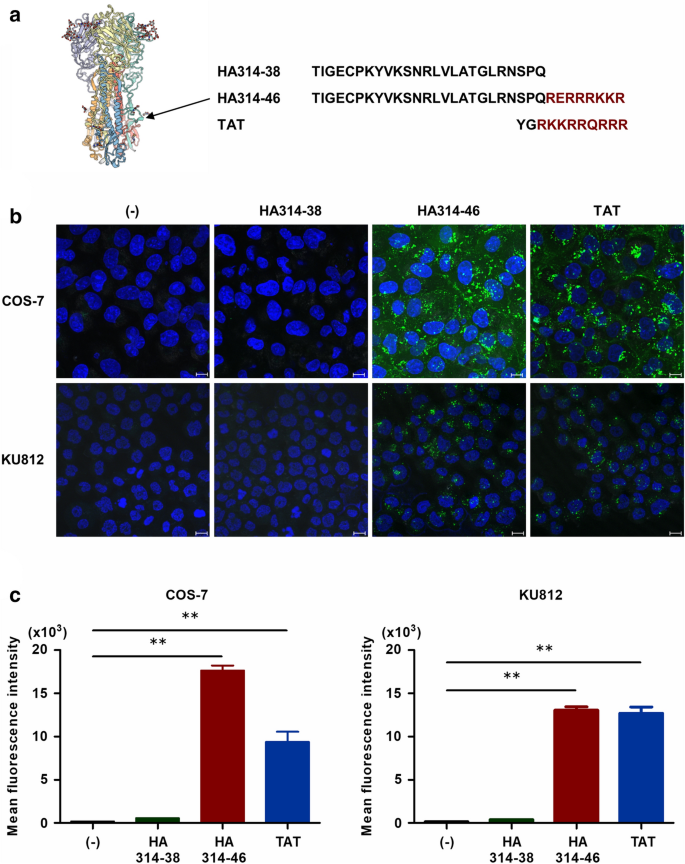
H5N1 HPAIV-derived HA314-46 peptide exhibits cell-penetrating activity. (a) Three-dimensional structure of HA protein and amino acid sequences of H5N1 HPAIV-derived HA314-38, HA314-46, and HIV-1-derived TAT peptides. The structure and sequence were obtained from the SWISS-MODEL Repository50 (https://swissmodel.expasy.org/repository/uniprot/D9I6N5) and the UniProt database (Protein ID; D9I6N5), respectively. Red-coloured residues represent multiple basic amino acids in HA314-46 and TAT peptides. (b,c) Cell-penetrating activity of HA314-46 peptide in COS-7 and KU812 cells. COS-7 and KU812 cells were incubated with 10 μg/mL of FITC-conjugated peptides at 37 °C for 60 min. Internalisation of the peptides was examined using confocal microscopy (b). Representative images from three experiments are shown. Green, peptide; Blue, nucleus. Scale bar, 10 μm. Mean fluorescence intensity (MFI) of FITC in viable cells was measured by flow cytometry (c). Data are presented as mean + SEM (n = 3). Asterisks indicate significant increase by one-way ANOVA with Bonferroni's multiple comparison test. **p < 0.01.
References
Katso myös OIE WAHIS AH5N1. Siin ei ole vielä merkattu tämän päivän FB tieto Israelin kalkkunoista.

Inga kommentarer:
Skicka en kommentar